


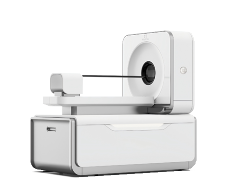
The small animal PET imaging system is a positron emission tomography imaging system equipped with independently developed submillimeter detectors. The system adopts high-precision crystal cutting technology, integrates low-power electronic systems, and has a globally leading spatial resolution. Can be used for imaging of single/multiple animals under different positron isotopes.
Product Features:
High resolution: The smallest crystal cutting in the industry, with a spatial resolution of less than 0.5mm, leading the world
High degree of freedom: expandable scanning field of view, aperture 130mm~290mm, axial field of view 26mm~158mm, freely selectable
High stability: The power of the detection system is less than 100W, and the detector does not require active cooling
100% independent intellectual property rights, independently producing core components
Application Cases:
Case 1: Mouse Brain Imaging
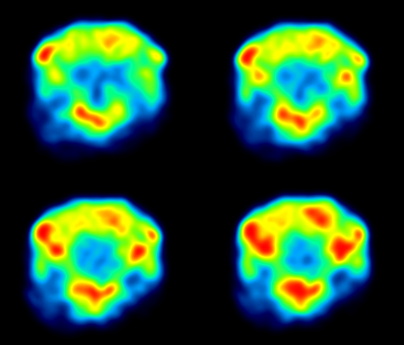
High resolution PET imaging of small animals provides More Details accurate information on brain function and molecular imaging, including key parameters such as neural metabolic activity in brain regions (such as glucose and neurotransmitter metabolism), receptor distribution, synaptic plasticity, and dynamic changes in neural circuits; The figure reflects the metabolic status of different brain regions in mice with different faults. Simultaneously possessing quantitative analysis capabilities, it can accurately reflect the degree of tracer enrichment in the brain through standardized uptake values (SUVs) and other methods. It has core value in studying the mechanisms of mouse brain diseases, such as amyloid deposition in Alzheimer's disease models, dopaminergic neuron damage in Parkinson's disease, abnormal discharges and metabolic remodeling in epilepsy, evaluating the efficacy of neuropharmaceuticals, such as the distribution and target validation of targeted receptor drugs in the brain, and analyzing molecular pathways of neural development and degeneration, such as synaptic development and dynamic tracking of neuroinflammation. In addition, the combination of multimodal imaging (such as PET/MRI fusion of brain anatomy and functional information) can promote the translation of basic neuroscience research into clinical practice (such as the validation of mouse models of novel neural tracers), providing key technical support for deciphering the mysteries of brain function and developing diagnosis and treatment strategies for brain diseases, and helping to achieve breakthroughs in cross scale research from "mouse brain mechanisms" to "human brain health".
Case 2: Mouse dynamic imaging
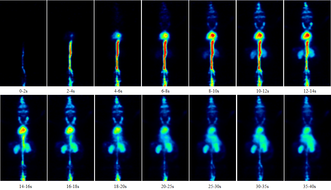
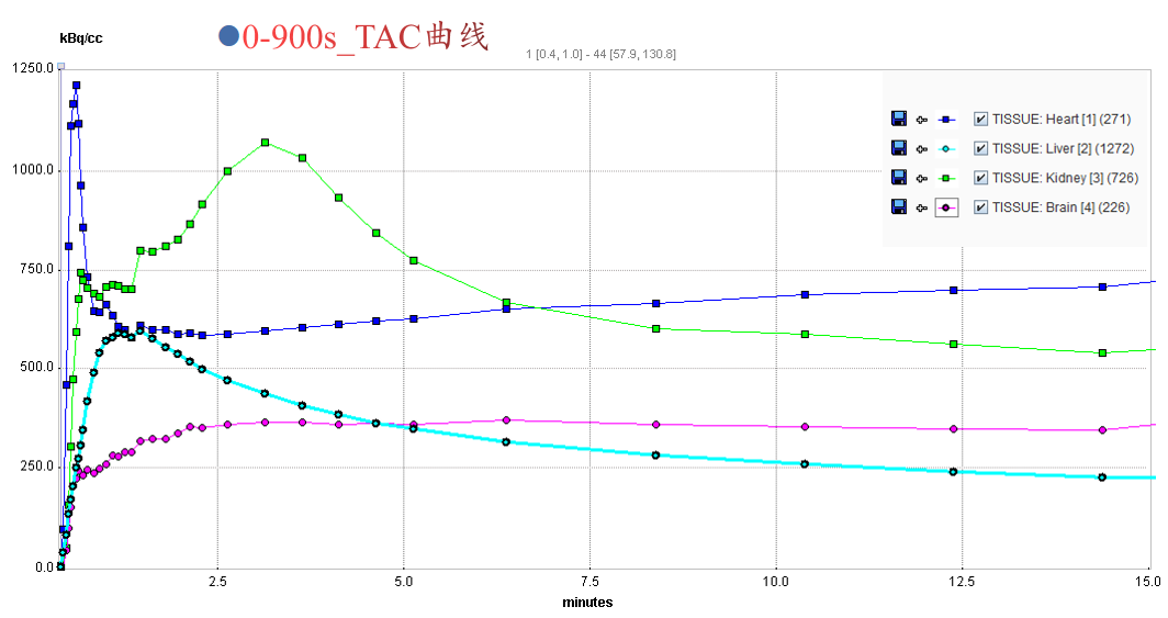
High resolution PET imaging of small animals provides More Details accurate dynamic functional and molecular distribution information, including the time concentration curves of tracers in mouse tissues (such as myocardium, brain, tumors, etc.) (such as metabolite uptake, drug distribution, and dynamic evolution of receptor binding), which can quantitatively analyze the spatiotemporal patterns of biological processes (such as metabolic heterogeneity of tumor growth, temporal dynamics of neurotransmitter transmission, and accumulation and clearance rates of drugs in target organs). The figure provides the process and information of the dynamic metabolic distribution of 0-900s 18F-FDG drug in mice. Its core value lies in the dynamic analysis of disease mechanisms, real-time monitoring of drug development, spatiotemporal tracking of gene and/or cell therapy, and precision in longitudinal research. In addition, by combining dynamic modeling (such as atrioventricular models and physiological pharmacokinetic models), small animal PET can convert image data into quantitative dynamic parameters, providing a mathematical and verifiable model basis for analyzing the spatiotemporal regulatory mechanisms of complex biological systems (such as functional connections of neural circuits and metabolic interactions in tumor microenvironments), promoting the upgrading of research paradigms from "static phenotype observation" to "dynamic mechanism analysis", and assisting in the deep expansion of mouse model research towards precise spatiotemporal biology.
Product Applications :
Tumor targeted imaging:
Suitable for evaluating the efficacy of FAP, PSMA and other target imaging agents in various tumor models.
Metabolic analysis of new drugs:
Suitable for dynamic monitoring of in vivo behaviors such as drug distribution, metabolism, and clearance.
Cardiovascular and cerebrovascular functional imaging:
Suitable for studying cardiac perfusion, cerebral blood flow, and functional activity in small animals.
Product Features:
High resolution: The smallest crystal cutting in the industry, with a spatial resolution of less than 0.5mm, leading the world
High degree of freedom: expandable scanning field of view, aperture 130mm~290mm, axial field of view 26mm~158mm, freely selectable
High stability: The power of the detection system is less than 100W, and the detector does not require active cooling
100% independent intellectual property rights, independently producing core components
Application Cases:
Case 1: Mouse Brain Imaging
High resolution PET imaging of small animals provides More Details accurate information on brain function and molecular imaging, including key parameters such as neural metabolic activity in brain regions (such as glucose and neurotransmitter metabolism), receptor distribution, synaptic plasticity, and dynamic changes in neural circuits; The figure reflects the metabolic status of different brain regions in mice with different faults. Simultaneously possessing quantitative analysis capabilities, it can accurately reflect the degree of tracer enrichment in the brain through standardized uptake values (SUVs) and other methods. It has core value in studying the mechanisms of mouse brain diseases, such as amyloid deposition in Alzheimer's disease models, dopaminergic neuron damage in Parkinson's disease, abnormal discharges and metabolic remodeling in epilepsy, evaluating the efficacy of neuropharmaceuticals, such as the distribution and target validation of targeted receptor drugs in the brain, and analyzing molecular pathways of neural development and degeneration, such as synaptic development and dynamic tracking of neuroinflammation. In addition, the combination of multimodal imaging (such as PET/MRI fusion of brain anatomy and functional information) can promote the translation of basic neuroscience research into clinical practice (such as the validation of mouse models of novel neural tracers), providing key technical support for deciphering the mysteries of brain function and developing diagnosis and treatment strategies for brain diseases, and helping to achieve breakthroughs in cross scale research from "mouse brain mechanisms" to "human brain health".
Case 2: Mouse dynamic imaging
High resolution PET imaging of small animals provides More Details accurate dynamic functional and molecular distribution information, including the time concentration curves of tracers in mouse tissues (such as myocardium, brain, tumors, etc.) (such as metabolite uptake, drug distribution, and dynamic evolution of receptor binding), which can quantitatively analyze the spatiotemporal patterns of biological processes (such as metabolic heterogeneity of tumor growth, temporal dynamics of neurotransmitter transmission, and accumulation and clearance rates of drugs in target organs). The figure provides the process and information of the dynamic metabolic distribution of 0-900s 18F-FDG drug in mice. Its core value lies in the dynamic analysis of disease mechanisms, real-time monitoring of drug development, spatiotemporal tracking of gene and/or cell therapy, and precision in longitudinal research. In addition, by combining dynamic modeling (such as atrioventricular models and physiological pharmacokinetic models), small animal PET can convert image data into quantitative dynamic parameters, providing a mathematical and verifiable model basis for analyzing the spatiotemporal regulatory mechanisms of complex biological systems (such as functional connections of neural circuits and metabolic interactions in tumor microenvironments), promoting the upgrading of research paradigms from "static phenotype observation" to "dynamic mechanism analysis", and assisting in the deep expansion of mouse model research towards precise spatiotemporal biology.
Product Applications :
Tumor targeted imaging:
Suitable for evaluating the efficacy of FAP, PSMA and other target imaging agents in various tumor models.
Metabolic analysis of new drugs:
Suitable for dynamic monitoring of in vivo behaviors such as drug distribution, metabolism, and clearance.
Cardiovascular and cerebrovascular functional imaging:
Suitable for studying cardiac perfusion, cerebral blood flow, and functional activity in small animals.


No.1 Gaochuang Road, Linyi Hi-Tech Electronics Industrial Park, Linyi City, Shandong Province, China
Room 801, Block B, Silicon Valley Building, South 7th Street, Shushan District, Hefei City,Anhui

Follow official account