


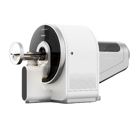
Small animal three mode imaging system is an integrated imaging system integrating three imaging modes of small animal PET, SPECT and CT. This system breaks through the limitations of a single imaging technology and integrates the distribution, metabolism, receptor expression, and other information of different nuclide labeled molecules with classical morphology, structure, density, and other information. It is currently one of the most economical and efficient complementary combinations.
Product Features:
High resolution PET system: the smallest crystal cutting in the industry, with a spatial resolution of less than 0.5mm, leading the world
High quality CT system: adjustable resolution CBCT, resolution ≤ 50um
High throughput SPECT system: Multiple mice are scanned simultaneously, with a single imaging field of view ≥ 80mm
High precision fused image: shared bed, position encoder, and high-precision image registration algorithm, precise image fusion registration
100% independent intellectual property rights, independently producing core components
Three mode integrated structure, personalized configuration PET/SPECT/CT
Application Cases:
Case 1: Multimodal imaging of myocardial infarction in yellow rats
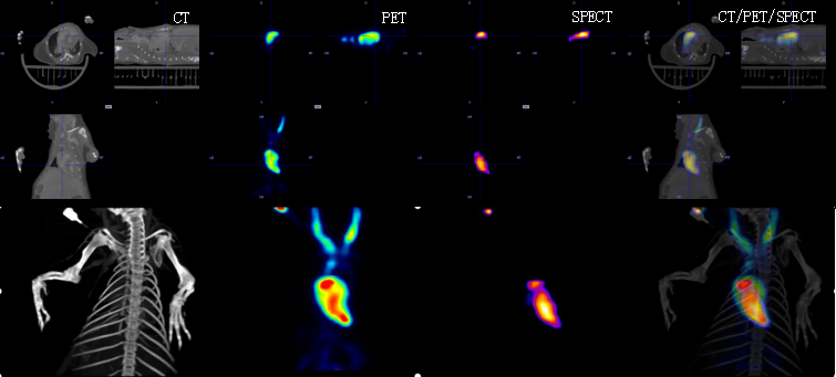
Small animal multimodal imaging (PET/SPECT/CT) provides multidimensional integrated information in the study of myocardial infarction in yellow rats, including: PET: quantifying myocardial glucose metabolism, perfusion imaging, and cell survival status through tracers such as 18F-FDG, Tc99 MIBI, CT iodofol, etc., to identify "metabolic perfusion mismatch" areas (surviving myocardium); SPECT: Dynamic evaluation of myocardial perfusion defect range and collateral circulation establishment using tracers such as Tc99 MIBI; CT: High resolution presentation of ventricular wall thickness, degree of cardiac chamber enlargement, and distribution of calcifications after myocardial infarction, accurately locating the anatomical boundary of the infarction.
Its core value lies in: multimodal collaborative analysis of pathological mechanisms/quantitative evaluation and precise classification/optimization of treatment strategies and efficacy monitoring/bridging role in translational medicine.
To sum up, PET/SPECT/CT multimodal imaging provides a full chain of technical support for mechanism analysis, efficacy evaluation, transformation and verification of myocardial infarction research in hamsters through the information integration of molecular function (PET), hemodynamics (SPECT) and anatomical structure (CT), breaks through the limitation of single mode, enables the paradigm upgrade of myocardial infarction pathology research from "fragmented observation" to "systematic analysis", and helps the innovation and implementation of accurate diagnosis and treatment strategies for ischemic heart disease.
Case 2: Tumor Targeted Imaging
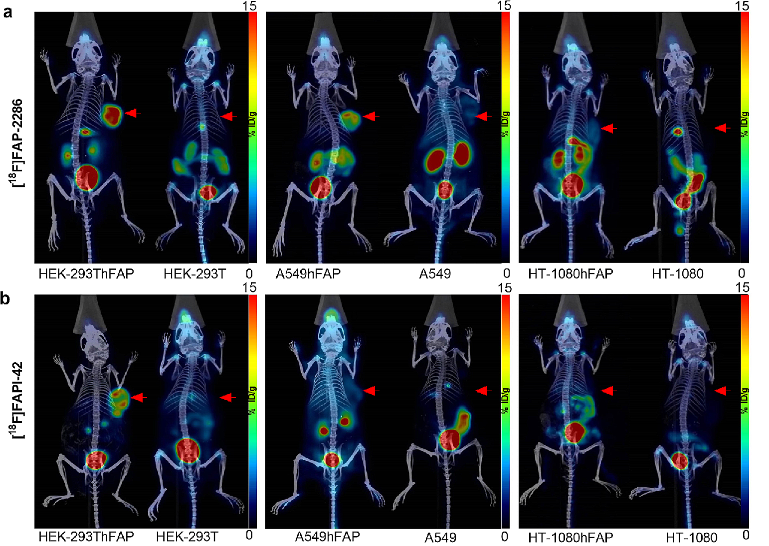
PET/SPECT/CT multimodal imaging, through the information coupling of molecular function (PET) - blood flow/receptor characteristics (SPECT) - anatomical structure (CT), provides full cycle technical support for tumor research, including mechanism analysis (from molecular phenotype to transfer path), efficacy evaluation (from metabolic response to structural regression), and transformation verification (from animal models to clinical programs), breaks through the limitation of single mode, enables the paradigm transformation of accurate tumor diagnosis and treatment from "empirical trial and error" to "data-driven design", and speeds up the pre clinical evaluation and transformation of innovative therapies.
The image shows that fibroblast activation protein (FAP) is highly expressed in many cancer associated fibroblasts (CAFs) of solid cancers, but is lowly or not expressed in normal tissues. The article used three novel FAP specific tracers to image six cell lines to validate the performance of [18F] FAP-2286;
The data was provided by the First Affiliated Hospital of Guangzhou Medical University and has been published in the European Journal of Molecular Imaging in Nuclear Medicine.
Product Applications:
Tumor targeted imaging:
Suitable for evaluating the efficacy of FAP, PSMA and other target imaging agents in various tumor models.
Metabolic analysis of new drugs:
Suitable for dynamic monitoring of in vivo behaviors such as drug distribution, metabolism, and clearance.
Cardiovascular and cerebrovascular functional imaging:
Suitable for studying cardiac perfusion, cerebral blood flow, and functional activity in small animals.
Bone metabolism research:
SPECT and CT fusion imaging can be used for modeling and evaluating skeletal system diseases.
Product Features:
High resolution PET system: the smallest crystal cutting in the industry, with a spatial resolution of less than 0.5mm, leading the world
High quality CT system: adjustable resolution CBCT, resolution ≤ 50um
High throughput SPECT system: Multiple mice are scanned simultaneously, with a single imaging field of view ≥ 80mm
High precision fused image: shared bed, position encoder, and high-precision image registration algorithm, precise image fusion registration
100% independent intellectual property rights, independently producing core components
Three mode integrated structure, personalized configuration PET/SPECT/CT
Application Cases:
Case 1: Multimodal imaging of myocardial infarction in yellow rats
Small animal multimodal imaging (PET/SPECT/CT) provides multidimensional integrated information in the study of myocardial infarction in yellow rats, including: PET: quantifying myocardial glucose metabolism, perfusion imaging, and cell survival status through tracers such as 18F-FDG, Tc99 MIBI, CT iodofol, etc., to identify "metabolic perfusion mismatch" areas (surviving myocardium); SPECT: Dynamic evaluation of myocardial perfusion defect range and collateral circulation establishment using tracers such as Tc99 MIBI; CT: High resolution presentation of ventricular wall thickness, degree of cardiac chamber enlargement, and distribution of calcifications after myocardial infarction, accurately locating the anatomical boundary of the infarction.
Its core value lies in: multimodal collaborative analysis of pathological mechanisms/quantitative evaluation and precise classification/optimization of treatment strategies and efficacy monitoring/bridging role in translational medicine.
To sum up, PET/SPECT/CT multimodal imaging provides a full chain of technical support for mechanism analysis, efficacy evaluation, transformation and verification of myocardial infarction research in hamsters through the information integration of molecular function (PET), hemodynamics (SPECT) and anatomical structure (CT), breaks through the limitation of single mode, enables the paradigm upgrade of myocardial infarction pathology research from "fragmented observation" to "systematic analysis", and helps the innovation and implementation of accurate diagnosis and treatment strategies for ischemic heart disease.
Case 2: Tumor Targeted Imaging
PET/SPECT/CT multimodal imaging, through the information coupling of molecular function (PET) - blood flow/receptor characteristics (SPECT) - anatomical structure (CT), provides full cycle technical support for tumor research, including mechanism analysis (from molecular phenotype to transfer path), efficacy evaluation (from metabolic response to structural regression), and transformation verification (from animal models to clinical programs), breaks through the limitation of single mode, enables the paradigm transformation of accurate tumor diagnosis and treatment from "empirical trial and error" to "data-driven design", and speeds up the pre clinical evaluation and transformation of innovative therapies.
The image shows that fibroblast activation protein (FAP) is highly expressed in many cancer associated fibroblasts (CAFs) of solid cancers, but is lowly or not expressed in normal tissues. The article used three novel FAP specific tracers to image six cell lines to validate the performance of [18F] FAP-2286;
The data was provided by the First Affiliated Hospital of Guangzhou Medical University and has been published in the European Journal of Molecular Imaging in Nuclear Medicine.
Product Applications:
Tumor targeted imaging:
Suitable for evaluating the efficacy of FAP, PSMA and other target imaging agents in various tumor models.
Metabolic analysis of new drugs:
Suitable for dynamic monitoring of in vivo behaviors such as drug distribution, metabolism, and clearance.
Cardiovascular and cerebrovascular functional imaging:
Suitable for studying cardiac perfusion, cerebral blood flow, and functional activity in small animals.
Bone metabolism research:
SPECT and CT fusion imaging can be used for modeling and evaluating skeletal system diseases.


No.1 Gaochuang Road, Linyi Hi-Tech Electronics Industrial Park, Linyi City, Shandong Province, China
Room 801, Block B, Silicon Valley Building, South 7th Street, Shushan District, Hefei City,Anhui

Follow official account